

articles
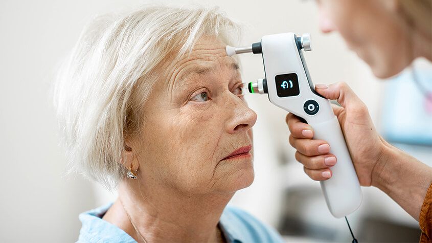
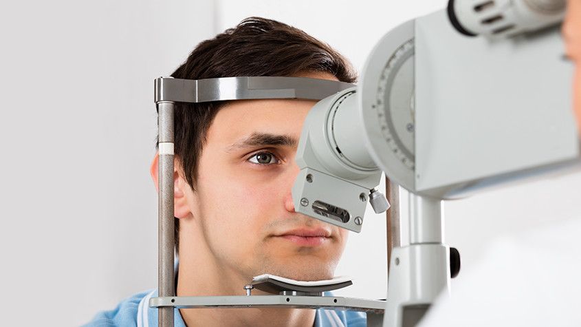
A tonometer refers to the equipment that is used in tonometry – a test that measures the pressure inside your eyes, also known as intraocular pressure or IOP for short. Tonometry is rarely performed at your average comprehensive eye exam unless you are at high risk of or have been already diagnosed with glaucoma. Fortunately, tonometry can be used to detect changes in eye pressure before they cause any symptoms, enabling prompt action to be taken before your vision is affected.
About glaucoma
Glaucoma is a common eye condition that occurs when the optic nerve, which connects the eye to the brain, becomes damaged. It’s normally caused by fluid building up in the front part of the eye, which causes the pressure inside the eyes to build. As the pressure increases, the optic nerve becomes increasingly damaged, and this prevents messages from being transmitted between your eyes and brain effectively. As a result, the patient’s vision becomes compromised. Without treatment, the level of vision loss will continue to increase. Unfortunately, any vision that has been lost as a result of glaucoma cannot be restored.
Most of the time, glaucoma develops very slowly which means that many people don’t realize that they are affected until some damage to their vision has already occurred. However, occasionally glaucoma can develop quickly, and symptoms do occur. These can include:
- Red eyes
- Intense headaches
- Tenderness around the eyes
- Eye pain
- Seeing rings/halos around lights
- Blurred vision
- Nausea and vomiting
If you notice any of these symptoms, it’s important that you make an appointment with your eye doctor right away so that you can be assessed. You are likely to have a tonometry test as part of this assessment.
What to expect from tonometry testing
There are various methods of tonometry testing, but many eye doctors use either Goldmann tonometry, which is the conventional technique to measure eye pressure, or electronic tonometry.
Goldmann tonometry testing is carried out using the Goldmann applanation tonometer, which is attached to a slit lamp microscope. This requires anesthetic eye drops to be used which numb your eyes, before a small probe is pressed gently against the eye, indenting the cornea. The pressure that the cornea pushes back onto the tonometer is what is measured to give your IOP reading. Electronic tonometry is where a handheld, mobile device is gently and quickly applied to the cornea to check the pressure, providing an accurate reading. Some eye doctors also offer non-contact tonometry which is where a puff of air is used to flatten the cornea, although this is reported to be less accurate than the Goldmann technique.
If you would like to find out more about Tonometry testing, please call our office to speak with our dedicated eyecare professionals.
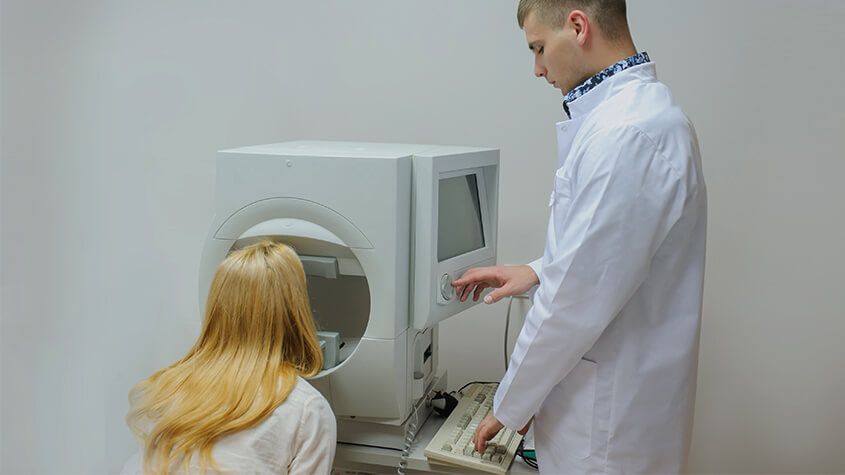
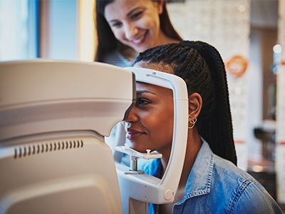
Visual field testing is an important part of most standard comprehensive eye exams. Also sometimes known as perimetry testing, Visual field testing is a method to measure the entire scope of vision of an individual, including their peripheral/side vision.
The importance of visual field testing
Visual field testing is one of the most effective diagnostic treatments in the detection of glaucoma. This is because when patients are affected by glaucoma, it is usually the peripheral vision that is affected by their condition first. However, it can also be used to detect central or peripheral retinal diseases, eyelid conditions such as drooping, optic nerve damage and conditions that affect the visual pathways from the optic nerve to the area of the brain where this information is processed into vision.
Visual field testing is also an important part of monitoring for people who are considered to be at risk for vision loss from disease and other problems, including those who have been diagnosed with the following:
- Multiple sclerosis
- Hyperthyroidism
- Pituitary gland disorders
- Central nervous system problems (such as a tumor that may be pressing on the brain)
- Stroke
- Diabetes
- High blood pressure
What to expect from visual field testing
There are a variety of methods that can be used to perform visual field testing, including:
Static automated perimetry. This is where a machine is used to quantify how well the patient is able to detect flashing lights of varying size and brightness in different areas of their visual field, while they concentrate on a central point. The patient responds by pushing a button when they see the light.
Kinetic perimetry. This involves points of light that are fixed in size and intensity and are presented along the patient’s peripheral vision, before being gradually moved inwards to determine their field of vision.
Visual field testing is non-invasive, painless and doesn’t require patients to have their eyes dilated. The results, which are usually presented in a series of charts, are digital and sent directly to your eye doctor for interpretation. Depending on the outcome of your results, you may be recommended for further diagnostic testing which could include blood tests. If you have been diagnosed with glaucoma, you will probably be recommended to have several visual field tests each year, which will help your eye doctor to monitor the progression of your condition and recommend treatments to slow it.
If you would like more information about visual field testing, or if you have concerns about your peripheral vision, please don’t hesitate to schedule an appointment with our experienced and knowledgeable eyecare team today.

Optomap is an innovative new technology that gives eye doctors the ability to perform ultra-wide retinal imaging that is far superior to what can currently be achieved using conventional retinal imaging options. In contrast to conventional retinal imaging, Optomap captures at least 50% more of the retina in a single capture, and with Optomap’s multi-capture function, up to 97% of the retina can be viewed. This gives eye care professionals greater opportunity to monitor the health and condition of patient vision.
Why is Optomap important?
Optomap is another great preventative eyecare technology tool. By allowing your eye doctor to have a comprehensive view of your retina, they will be able to detect any developing eye diseases early on, before they have a detrimental impact on your vision and day to day life. Not only can Optomap detect eye conditions such as retinal holes, retinal detachment, macular degeneration and diabetic retinopathy, but it can also be used to identify some general health conditions such as cardiovascular disease, stroke and cancer.
What to expect from Optomap scanning
Optomap is a fast, painless and non-invasive procedure that is suitable for patients of all ages, even children and pregnant women. Many patients require their eyes to be dilated ahead of the scan and will be given eyedrops which will widen their pupils and make it easier for the camera to see the structures inside the eye. Pupil dilation is painless, but patients may feel more sensitive to light both during their Optomap scan and afterwards for up to 24 hours. You may also have slightly blurred vision for a few hours. Once your eyes are dilated, you’ll be sat down and asked to look into a small device that will take the pictures of your retina. A short flash of light will let you know that the image has been taken, and the entire imaging is over in just a few seconds. The results will be sent digitally to your eye doctor who will then evaluate them. The results will also be stored on your personal optical record for future information.
If you would like more information about what is involved in Optomap, or to schedule an appointment for this effective screening technology, please contact our eyecare team.
We all want to look our best and in the last decade, we have seen a significant increase in the number of people seeking cosmetic services in order to enhance their appearance. With our eyes being our most distinguishing feature, we want to make the most of them. Thankfully there is now a range of cosmetic services that can help to rejuvenate our eyes and the area around them to keep them fresh, young and wrinkle-free.
Let’s take a look at some of the services on offer.
Pigment removal
The brown pigment spots that appear on the face are often referred to as age spots and are a result of sun exposure. With age, the repeated exposure to UV rays causes melanin, a compound that is responsible for pigmentation and protecting the skin begins to clump together to form an area of hyperpigmentation. Whilst they aren’t any cause for concern, many people feel that they look unsightly. Luckily, there are a number of different treatments that you can get to remove them including topical creams, laser therapy, and chemical peels.
If you are suffering from darker pigmentation then we strongly recommend that you make an appointment with a qualified dermatologist who will recommend the best course of treatment for you, based on your specific needs.
If you are one of the thousands of people considering LASIK laser eye surgery, then you will probably be gathering as much information as possible about the treatment. By this point, you are probably aware of the benefits that LASIK offers, such as a reduced or eliminated need for glasses or contact lenses and greater convenience in your day to day life. However, for many patients, despite the advantages of LASIK, the thought of surgery on their eyes is still a cause of anxiety and fear. One of the best ways to alleviate this concern is to find out more about what the procedure entails.
Your consultation
Before you can be approved for any form of laser vision correction, including LASIK, you will need to attend a consultation appointment with your surgeon. During the consultation, he will perform an examination of your eyes and use your medical and ocular history to determine if you are a good candidate for the procedure. He will also speak to you about the expected outcome from your surgery, making you aware that while LASIK will dramatically improve your eyesight, there is no guarantee that you will not need to wear glasses in some situations, such as while driving in the dark.
How LASIK Works
LASIK uses a cool, ultraviolet beam of light to reshape the patient’s cornea. Doing so will more accurately focus the light that enters the eye on to the retina, thus improving the patient’s vision. The way in which the cornea needs to be reshaped will depend on the visual needs of the patient. For example, a patient who is far-sighted will need their cornea reshaping to be steeper to experience better eyesight. Alternatively, a patient who is near-sighted will require their cornea to be flattened in order to improve their vision. LASIK can also smooth an irregular cornea into a more standard shape, meaning that the procedure can also be used to correct astigmatism.
The LASIK procedure
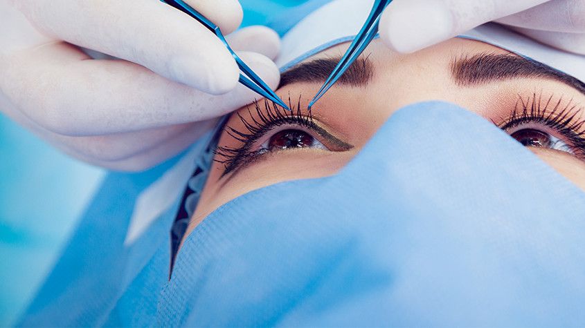
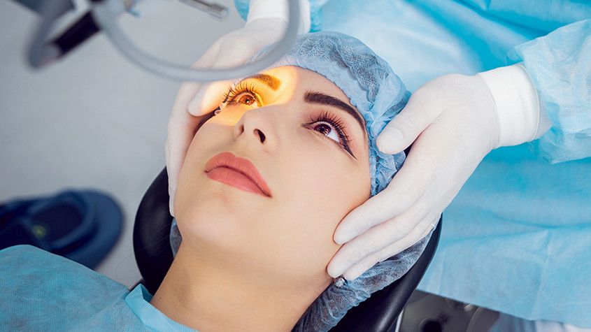
The LASIK procedure is very fast and straightforward. Although you will probably be in the surgical suite for around half an hour, the actual process only takes a couple of minutes per eye. The rest of the time will be spent preparing and ensuring that you are comfortable. Anesthetic eye drops are given to patients before their procedure so that the entire process is pain-free. If you are particularly anxious, it may also be possible for you to be slightly sedated – this should be discussed with your doctor at your consultation appointment.
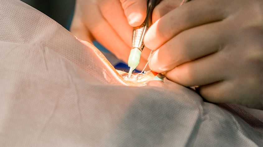
Once you are in position, we will use a femtosecond laser to cut a thin, circular flap into the outer cornea. This can then be pulled back to reveal the underlying corneal tissue, known as the stroma so that it can be reshaped using the laser. The exact path that the laser needs to take, known as the topography, will have been pre-programmed ahead of the procedure and can be followed with complete precision and accuracy.
Once the reshaping is complete, the flap is replaced back over the eye and the surgery is complete. There is no need for sutures or bandages as the cornea will start to heal immediately and without any medical intervention.

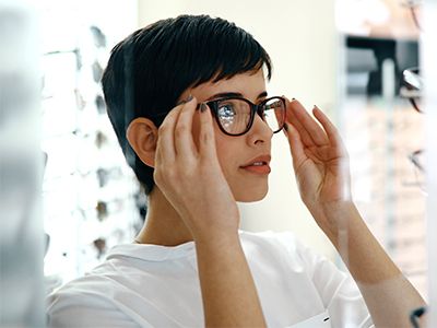
If you already rely on wearing glasses or contact lenses to be able to see clearly, you may be frustrated with the effect that they have on your life. Regular vision tests, finding glasses to suit your face shape, having to remember to take eyeglasses with you wherever you go, prescription sunglasses, fiddly contact lenses… the list of inconveniences associated with conventional ocular solutions is extensive.
LASIK is a modern, minimally-invasive procedure that can substantially reduce or eliminate your need to use eyeglasses or contact lenses, allowing you to enjoy life without limitations or inconvenience. The popularity and success of LASIK laser eye surgery have helped to make it the number one elective surgery across the globe.
Candidacy for LASIK
LASIK has an extremely high success rate. According to the American Society of Cataract and Refractive Surgery, 96% of patients achieve 20/20 vision or better. However, it’s high success rate doesn’t make LASIK automatically the right solution for everyone.
Candidacy for LASIK is assessed by our doctors on a case by case basis so that you be certain that whatever treatment is recommended for you, it will give you the very best opportunity to improve your vision. During your consultation, our doctors will perform a thorough examination of your eyes and vision, ask you about your general health and talk you through both the procedure and aftercare.
The general guidelines for LASIK candidacy state that patients must:
be at least 18 years of age
have had stable vision with no prescription changes for a minimum of 12 months
have a current prescription for eyeglasses or contact lenses that falls between specified parameters (Our doctors will be aware of what these parameters are)
have no significant medical or eye-related problems such as glaucoma, macular degeneration or diabetic retinopathy
have no history of corneal disease
not be pregnant or nursing at the time of the procedure
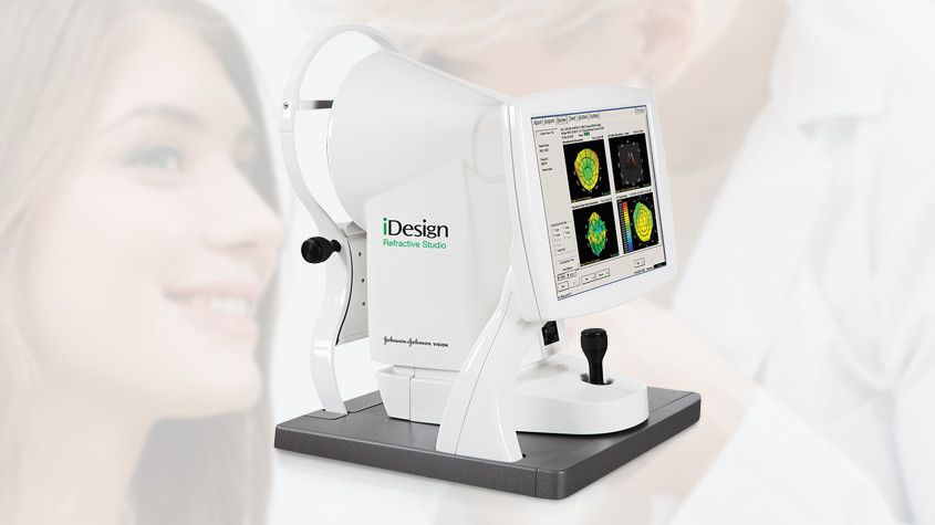
If you thought that just laser eye surgery was impressive, then be prepared to be astounded by the arrival of iDesign custom Lasik. iDesign is the most advanced wavefront-guided laser treatment available. The system creates a uniquely accurate measurement of the entire optical system that can be relied upon completely without any guesswork. This is then used to create a highly detailed topography or map of your eye, making it easier for your surgeon to pinpoint the areas that need addressing with pinpoint precision.
How accurate is iDesign?
iDesign is extremely accurate due to its diagnostic equipment that is able to determine 1,257 different micro eye prescriptions. Each one of these will measure the eye prescription to 0.01 of a dioptre strength – 25 times more than what a regular prescription for glasses or contact lens prescription is measured to.
Who is iDesign suitable for?
While iDesign Custom Lasik can benefit almost any patient with refractive problems, it is particularly beneficial for people who have unusual corneas or have had eye surgery in the past.
Is it painful?
Laser eye surgery is generally considered a pain-free procedure by the majority of patients. Anesthetic eye-drops are used to numb the outer area of the eye during the course of the surgery. Once these have worn off, some patients do experience some mild discomfort during the days following the procedure, but over the counter pain relief is usually sufficient to relieve this.
Astigmatism is a relatively common eye disorder that causes the vision to be blurred or distorted. It occurs when the lens part of the eye, known as the cornea, isn’t perfectly curved and instead resembles a football rather than a soccer ball. This means that the light entering the eye comes through at a distorted angle, making the object appear blurry and out of focus. There are several ways in which it is possible to treat astigmatism, including laser eye surgery and corrective lenses. However, another possibility is a solution referred to as limbal relaxing incisions.
What are limbal relaxing incisions?
Limbal relaxing incisions are microscopic cuts to an area in the eye known as the limbus. This helps to relax the curve in the cornea and improve its ability to focus light correctly. It can significantly improve your astigmatism and the overall quality of your vision.
Am I a good candidate for limbal relaxing incisions?
If you have astigmatism, are over 18, in good general health and have no major eye conditions, then chances are you are a good candidate for limbal relaxing incisions. Make an appointment with your eye doctor to discuss your candidacy further.
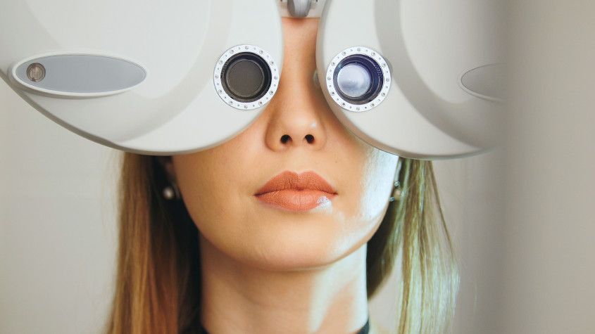
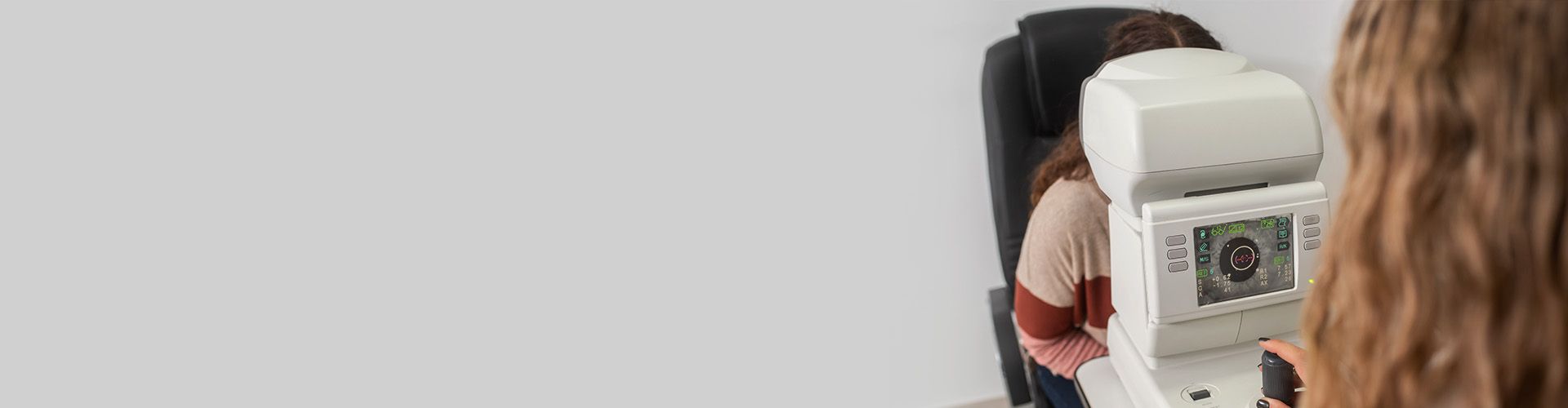
A refraction test, also called a vision test, is usually performed as a part of a routine eye examination. The purpose of this test is to determine if a person has a refractive error which would then mean the patient would need glasses or contact lenses.
What Is The Normal Value for Refraction Test?
A value of 20/20 is normal (optimum) vision. This means that individuals who have 20/20 vision are able to read letters that are 3/8-inch (1 centimeter) tall from 20 feet (6 meters) away. The normal uncorrected vision (without glasses or contact lenses) refractive error is zero (plano). Individuals who don’t have 20/20 vision, have what is called a refractive error. A refractive error means that the light is not bending properly when it passes through the lens of the eye. The refraction test will tell the doctor what prescription lens should be used in order to have 20/20 vision.
For people over age 40 who have normal distance vision but difficulty with near vision, a refraction test with a small type size is used to determine normal near vision and the correct power of reading glasses.
How Is The Refraction Test Performed?
The test is performed by having the patient seated in a chair that has a special device (called a phoropter or refractor) attached to it. The patient looks through the device and focuses on an eye chart 20 feet (6 meters) away. The device contains lenses of different strengths that can be moved into the patient’s view. The test is performed one eye at a time. If the patient is wearing contact lenses, they should be removed before the test.
In case the final vision is less than 20/20 even with lenses, then there is probably another non-optical problem with the eye. The vision level achieved during the refraction test is called the best-corrected visual acuity (BCVA).
What Are The Causes of Abnormal Refraction Test Results?
Abnormal results may be due to:
Astigmatism (abnormally curved cornea causing blurred vision)
Hyperopia (farsightedness)
Myopia (nearsightedness)
Presbyopia (inability to focus on near objects that develop with age)
Other conditions under which the test may be performed:
Corneal ulcers and infections
Loss of sharp vision due to macular degeneration
Retinal detachment (separation of the light-sensitive membrane (retina) in the back of the eye from its supporting layers)
Retinal vessel occlusion (blockage in a small artery that carries blood to the retina)
Retinitis pigmentosa (an inherited disorder of the retina)
There is an art to refraction and the optometrist will always answer the patient’s questions and as well as discuss their findings. Based on the results of the refraction test, they can determine the amount of myopia, hyperopia or astigmatism.
The parts of a comprehensive eye examination vary according to the patient's age, date of last exam, and other factors. Not all parts of the eye exam may be needed or performed, but the first part of the eye exam will include documenting medical history. Here are some eye and vision tests that are likely to be encountered during a comprehensive eye exam:
Visual Acuity Tests
Visual acuity tests measure the sharpness of vision and are usually performed using a projected eye chart to measure the distance visual acuity and a hand-held small acuity chart to measure the near vision (for reading).
Color Blindness Test
A screening test that checks the color vision is often performed early in a comprehensive eye exam to rule out color blindness.
Cover test to check eye alignment.
A test used to assess strabismus or a more subtle binocular vision problem that could cause eye strain or amblyopia (lazy eye).
Ocular Motility (Eye Movements) Testing
Ocular motility testing is performed to determine how well eyes can follow a moving object and/or quickly move between and accurately fixate on two separate targets.
Stereopsis (Depth Perception) Test
This is used to test perception of depth and 3-dimensional structure obtained on the basis of visual information deriving from two eyes by individuals with normally developed binocular vision.
Retinoscopy
This test is used to estimate which lens powers will best correct distance vision. Based on the way the light reflects from the eye, the doctor is able to obtain an approximation of the eyeglass prescription. This test is useful for children and patients who are unable to accurately answer the doctor's questions.
Manual refraction with a phoropter.
This is the test used to determine the exact eyeglass prescription.

© 2024 PersonalEyes Vision Care. All rights Reserved. Accessibility Statement | Privacy Policy | Sitemap

Powered by: